
Diagnosis
Can imaging be negative and still help plan endometriosis surgery?
The short answer
Yes, imaging can be negative and still help plan endometriosis surgery. Small or superficial disease may not show up, but imaging can help map deeper disease and assess whether nearby organs may be involved, helping plan the surgical team and anticipate complexity.
Yes, imaging can be negative and still be very useful for planning surgery. Small or superficial peritoneal endometriosis may not show up on ultrasound or MRI, so a normal report doesn’t rule out disease.
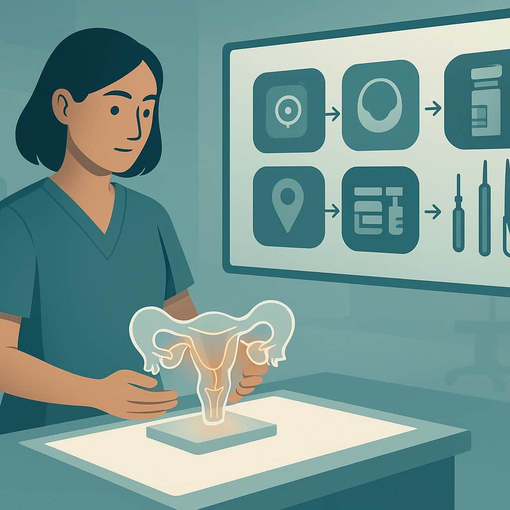
Even when it doesn’t show every lesion, imaging can help identify or map deeper disease, ovarian endometriomas, adhesions, and whether nearby organs may be involved. That information helps us plan the right surgical team, anticipate complexity, and have a clear, informed consent discussion before the procedure. When imaging and symptoms don’t match, our surgical assessment and intraoperative findings still guide what we treat. We’re happy to review your images and history with you in a consultation.
Where to go from here
Understand it, compare it with what you're feeling, then talk it through with us.
Step 2 · Compare
Symptoms it can relate to
Step 3 · Speak with Us
Imaging Normal? Surgery Still Possible
Normal imaging doesn’t rule out endometriosis. Our specialists use all info to plan precise, effective surgery tailored to you.
Questions people ask next
All diagnosis questionsCan imaging predict if I’ll need bowel, bladder, or ureter surgery?
Yes, high-quality pelvic ultrasound and MRI can often help anticipate whether bowel, bladder, or ureter surgery may be needed. Imaging helps plan the safest approach, but the definitive assessment and exact surgical plan are confirmed during surgery.
Do I need imaging or special prep before endometriosis excision?
In many cases, yes: targeted transvaginal ultrasound and/or pelvic MRI can help plan endometriosis excision more safely and thoroughly. Prep depends on imaging and symptoms; some patients may need bowel preparation or temporary ureteral stents.
Why was more endometriosis found in surgery than on imaging?
Surgery can reveal more endometriosis because scans may miss lesions that are small, superficial or hidden by normal anatomy. Imaging helps with surgical planning, but it can’t always show the full extent of disease or adhesions seen during laparoscopy.
When is imaging most helpful for endometriosis?
Imaging is most helpful when clarifying what may be driving your symptoms before treatment, or when we suspect deeper disease that may involve the bowel or bladder. It can help guide surgical planning, but it can’t rule out endometriosis.
Further reading
Do You Need Two Ultrasounds Before Bowel Endometriosis Surgery?
How TVS (transvaginal) and ERUS (endorectal) map rectal endometriosis, guide bowel surgery planning, flag stenosis and risks, and who benefits.
Deep Infiltrating Endometriosis: Current and Future Treatments
Deep infiltrating endometriosis: symptoms, causes, diagnosis and treatment. Medical therapy, ICG-guided surgery, stenting, pathology, and future outlook.
Gynecologic Oncologists - Safest Choice for Complex Endometriosis Surgery?
For deep or advanced endometriosis, oncology-trained surgeons often provide safer, more complete excision. Learn what this means for you at the Lotus Endometriosis Institute.