

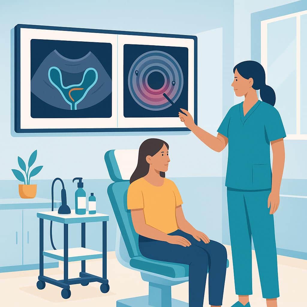
Do You Need Two Ultrasounds Before Bowel Endometriosis Surgery?
How TVS (transvaginal) and ERUS (endorectal) map rectal endometriosis, guide bowel surgery planning, flag stenosis and risks, and who benefits.
8 minute read
Read moreDiscover how Lotus can guide you toward lasting relief.
Explore why patients choose Lotus
Search expert-written answers, browse by topic, or find information based on where you are in your journey.
Explore All KnowledgeReach out and start your healing journey.
Schedule an AppointmentPre- and intraoperative imaging to map disease and guide precise excision—ultrasound, MRI, and fluorescence guidance—enhancing surgical planning, protecting vital structures, and lowering recurrence in deep and complex endometriosis.
Imaging for surgery focuses on mapping endometriosis before and during an operation so the team can plan precise, organ‑sparing treatment. High‑resolution, expert transvaginal or transrectal scans characterize deep nodules, tethering, and the “sliding sign,” while MRI outlines multi‑compartment disease and nearby structures, including bowel, bladder, ureters, and nerves. This planning helps select the right approach, assemble colorectal or urology support when needed, anticipate adhesions, and protect fertility and function. It differs from diagnostic imaging by prioritizing operative decisions and risk reduction rather than simply confirming disease. See Ultrasound and MRI for detection details.
In the operating room, near‑infrared fluorescence with indocyanine green can clarify ureter location and confirm blood flow after bowel or ureter resections, lowering complications like leaks or ischemia. When imaging and surgical strategy align, complete Excision Surgery becomes more feasible, recurrence risk falls, and recovery is safer. Guidance here also clarifies who benefits most—especially those with suspected Deep Infiltrating Endometriosis—and how results influence single versus staged Laparoscopy and specialist involvement.
Deep infiltrating endometriosis (DIE) is endometriosis that grows deeper into tissue, often described as more than ~5 mm below the surface. It commonly involves structures like the uterosacral ligaments, rectovaginal space, bowel, bladder, or ureters. Because it can inflame, scar, tether, or even narrow or obstruct nearby organs, DIE may cause non-gynecologic symptoms such as bowel or urinary pain, painful sex, nerve-type pelvic pain, or symptoms that don’t match a routine pelvic exam.
Treatment depends on where the disease is and what it’s affecting, but DIE is the subtype most likely to require specialized surgical planning. When surgery is appropriate, meticulous excision (removing disease at its roots rather than burning the surface) is the gold-standard approach for durable symptom relief and addressing organ involvement. In complex cases, this can include careful work around the bowel, bladder, and ureters. Our team focuses on advanced, precision excision, often using robotic technology, with a tailored plan that prioritizes safety, completeness, and your goals, whether that’s pain relief, fertility, or protecting organ function.
Because DIE can be missed on basic exams and even normal imaging, evaluation often requires a higher index of suspicion and the right strategy for mapping disease before any procedure. If DIE sounds like it could explain your symptoms, explore our detailed resources on deep disease and excision, or reach out to schedule a consultation so we can review your history, imaging, and next steps together.
The Enzian score is a detailed way for clinicians to describe where deep infiltrating endometriosis (DIE) is located and how extensive it is. Unlike simple staging systems, Enzian focuses on endometriosis that grows into deeper tissues and can involve structures like the uterosacral ligaments, rectovaginal area, bowel, bladder, and ureters. These areas often drive bowel, urinary, or deep pain symptoms.
In practice, an Enzian classification helps your surgical team communicate the anatomic pattern of disease and plan the right imaging, operative approach, and multidisciplinary support when organs may be involved. It’s also a reminder that symptom severity doesn’t always match what’s seen on exam or imaging: deep disease can be easy to miss without a targeted evaluation. If you’ve been told your findings are “mild” but your symptoms suggest deeper involvement, our team can help interpret prior reports and discuss what an Enzian-style mapping and excision-focused plan could look like.
It’s very common for surgery to reveal more endometriosis than ultrasound or MRI suggested. Imaging is best viewed as a tool to estimate likelihood and to map certain higher-risk areas for surgical planning, rather than as a reliable yes/no detector for every lesion. Many endometriosis lesions are hard to see on scans because they can be small, superficial, hidden by normal anatomy, or located in areas where imaging performance varies and interpretation depends heavily on technique and experience.
Scans are also better at identifying some patterns, like ovarian endometriomas or certain deep bowel disease, than at detecting disease on ligaments, in the bladder/anterior compartment, or in complex multi-compartment cases. Imaging also can’t always capture the full extent of adhesions, scar-like tissue, or subtle inflammatory changes that may become obvious only when the pelvis is directly inspected during laparoscopy.
When we plan surgery, we consider imaging alongside your symptom history, exam findings, and overall pattern. The goal is safe, complete mapping and excision when appropriate. If your operative findings didn’t match your scan, it doesn’t mean the imaging was pointless or that your symptoms were exaggerated. It usually reflects the known limits of what scans can show. If you’re trying to make sense of your results or next steps, our team can help you review what was found, what was removed, and what else (like adenomyosis or coexisting pain drivers) may still need to be addressed.
Usually, no bowel prep is needed for a standard transvaginal pelvic ultrasound used to evaluate suspected endometriosis. Most patients can eat, drink, and take medications as normal unless the imaging center gives you different instructions. The key is to arrive with the bladder filling they request.
Bowel prep is more commonly discussed when we’re specifically trying to map suspected bowel deep endometriosis (especially rectal involvement) with specialized imaging like an endorectal ultrasound, or when a radiology team uses a particular protocol to improve visibility of the bowel wall and surrounding tissues. If your symptoms suggest bowel involvement, our team focuses on choosing the right type of imaging and the right interpretation so the results help guide next steps.
If you’re unsure what test you’re scheduled for, reach out to confirm whether it’s a routine transvaginal ultrasound or a bowel-focused study and what preparation, if any, is expected. We can also help you decide whether additional mapping would be useful based on your symptom pattern and exam findings.
“Restricted sliding” on a pelvic ultrasound usually refers to a limited “sliding sign”: nearby pelvic structures don’t glide smoothly against each other when gentle pressure is applied with the ultrasound probe. In a typical pelvis, organs like the uterus, ovaries, bowel, and the space behind the uterus (often called the pouch of Douglas) should move freely relative to one another.
When sliding is restricted, it can suggest adhesions (scar tissue) or deep endometriosis tethering tissues together, sometimes described as fixed anatomy. It’s not a diagnosis by itself and doesn’t tell us the full extent of disease, but it’s a meaningful clue that can guide next-step imaging and, if surgery is being considered, preoperative planning. If your report mentions restricted sliding alongside symptoms like deep pelvic pain, painful sex, pain with bowel movements, or cyclical bowel/bladder flares, our team can help interpret what that combination may mean in your specific case and which evaluations are most useful next.
On pelvic MRI, “T2 dark plaque” describes an area that looks dark on the T2 sequence (a common MRI setting that highlights fluid and soft-tissue differences). Radiologists often use this phrase when they see a plaque-like region of low T2 signal that suggests dense, fibrotic tissue, often a scar-like change, rather than a simple fluid-filled cyst. In endometriosis, low T2 signal plaques can be seen with deep infiltrating disease or adhesions, because chronic inflammation can lead to fibrosis that looks dark on T2.
That said, “T2 dark plaque” is a descriptive imaging term, not a diagnosis by itself. Its meaning depends on the exact location, for example, behind the uterus, along the uterosacral ligaments, or near the bowel or bladder, and whether other supportive MRI features are present, since some benign non-endometriosis processes can also look T2-dark. If your report mentions a T2 dark plaque and you have symptoms that fit organ involvement (bowel, bladder, deep sex pain, severe cyclical pelvic pain), our team can review your imaging and history together and help you understand whether the finding is likely clinically significant and how it may affect next steps in treatment planning.
Sometimes, especially if imaging or exam suggests deeper rectal or sigmoid involvement and there is a real possibility that the bowel wall may need to be opened, repaired, or resected. Bowel endometriosis can range from superficial implants on the outer surface of the bowel to deep disease that infiltrates the muscular layers and narrows or distorts the lumen. The deeper it goes, the more important it is to have a team that can safely handle bowel entry, suturing, stapling, and anastomosis if needed. In most cases, a general surgeon or colorectal surgeon is required, unless your primary surgeon is a gynecologic oncologist or your team includes one. All three are credentialed in bowel surgery.
In many patients, bowel-type symptoms come from inflammatory pelvic disease and adhesions even without full-thickness bowel involvement, and surgery may be limited to careful excision off the bowel surface, or “shaving,” without requiring a formal bowel procedure. Ideally, the decision is made with pre-op mapping (often ultrasound and/or MRI) and a plan that matches your anatomy, so you’re not surprised in the operating room by an unanticipated higher-risk bowel step.
In our practice, we have a unique offering: Dr. Vasilev is a gynecologic oncologist who is trained and credentialed to perform bowel surgery. We plan bowel endometriosis surgery around safety and completeness, using robotic excision for precision, and we coordinate the right surgical partners when the imaging, exam, or history suggests a disc excision or segmental resection could be needed. For example, there may be evidence of other disease like diverticulosis or other bowel findings. If you tell us your symptoms and what your imaging shows (or if you’re unsure), our team can help you understand whether a colorectal surgeon should be involved from the start and what that means for recovery and outcomes.
Follow-up imaging doesn’t follow a single fixed schedule. It’s individualized based on what we’re monitoring. We use ultrasound and/or MRI most often when it will change decisions, such as tracking ovarian endometriomas, mapping suspected deep disease (bowel, bladder, or uterosacral involvement), or evaluating adenomyosis or a new pelvic mass. Symptoms and imaging don’t always match, so the goal is to get the right test at the right moment rather than scan on a timer.
In many cases, a practical framework is a baseline post-op check once initial healing is complete (often around 6–12 weeks), a planned reassessment around 6–12 months, and then annual follow-ups, especially if you had endometriomas, deep infiltrating disease, or persistent symptoms. Imaging may not be needed at every visit, but we’re more likely to recommend it when symptoms change, when prior imaging was incomplete, or when we need a clearer multi-compartment map to guide next steps. If you share your prior imaging reports, along with images if available, our team can tell you what’s worth repeating, what’s not, and how often surveillance makes sense for your specific pattern and goals.
How TVS (transvaginal) and ERUS (endorectal) map rectal endometriosis, guide bowel surgery planning, flag stenosis and risks, and who benefits.
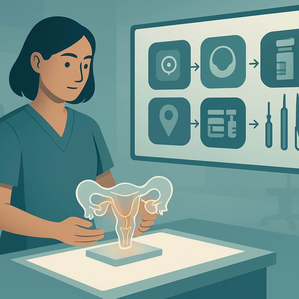
Deep infiltrating endometriosis: symptoms, causes, diagnosis and treatment. Medical therapy, ICG-guided surgery, stenting, pathology, and future outlook.
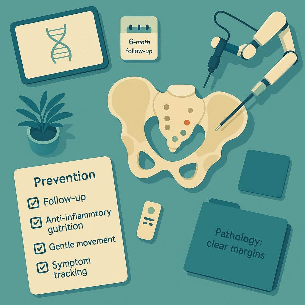
Learn why endometriosis recurs—incomplete excision, hormonal, immune, toxin and molecular factors—and how precise robotic surgery and 3D optics can reduce risk.
Lotus Endometriosis Institute provides California-based surgical evaluation and advanced excision care for patients with suspected endometriosis, adenomyosis, complex pelvic pain, and related conditions.
Many patients contact us from outside California to learn whether traveling for in-person evaluation and possible surgery may be appropriate.
2121 Santa Monica Blvd, Santa Monica, CA 90404
8am - 5pm
Monday - Friday
154 Traffic Way, Arroyo Grande, CA 93420