
Deep Disease
What tests diagnose rectovaginal septum endometriosis?
The short answer
Expertly performed transvaginal ultrasound and pelvic MRI can help map suspected rectovaginal septum endometriosis, but normal imaging can’t reliably rule it out. Surgery with biopsy is the only way to definitively confirm endometriosis.
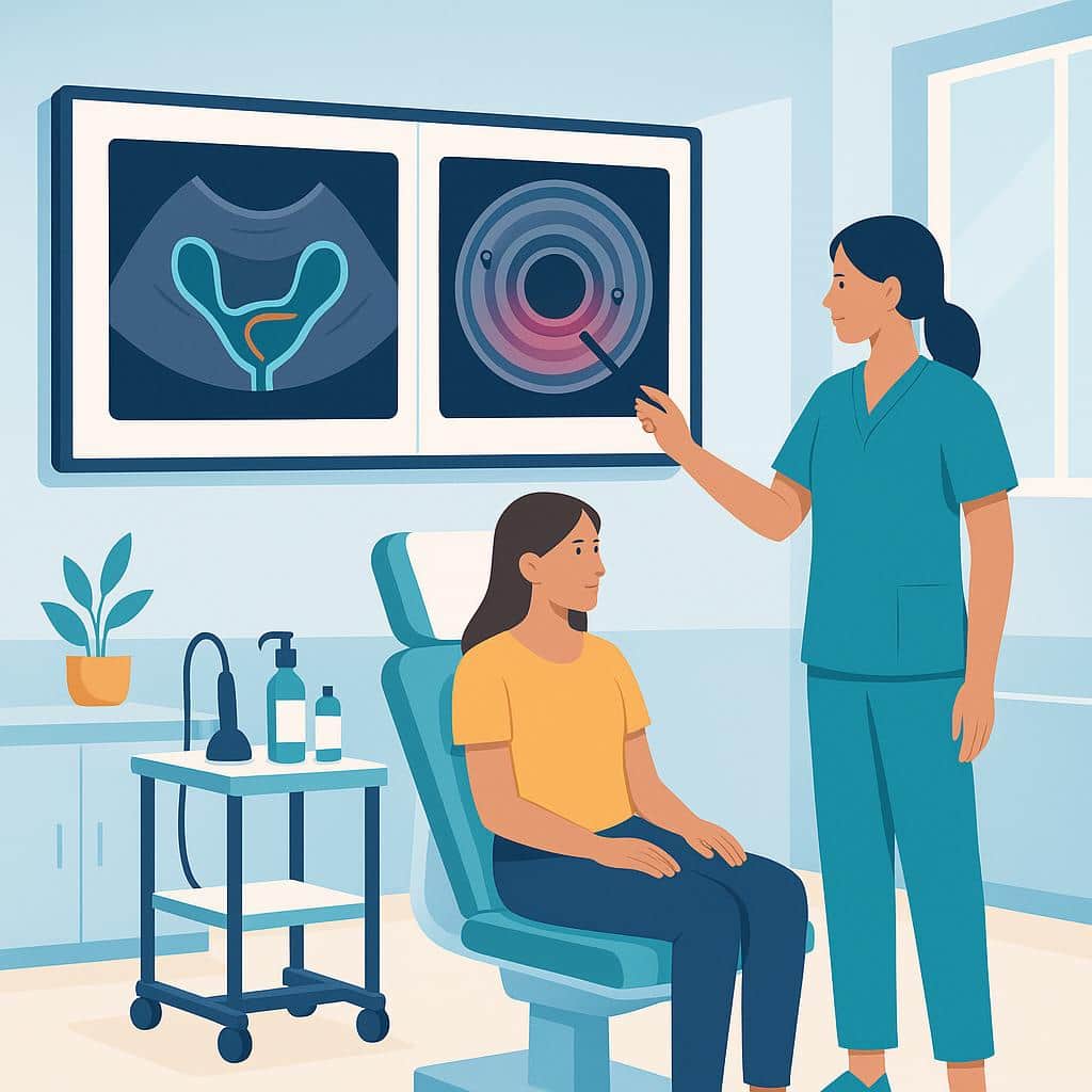
Rectovaginal septum endometriosis is usually evaluated through your symptom history, a targeted pelvic exam, and imaging interpreted by clinicians who routinely look for deep endometriosis. On exam, we often look for deep tenderness, thickening, or a firm nodule behind the vagina and correlate those findings with symptoms like deep pain with sex, pain with bowel movements, and cyclical rectal or pelvic pressure.
The most useful tests are expertly performed transvaginal ultrasound and pelvic MRI. These can help map suspected deep disease and plan surgery, but normal imaging still can’t reliably rule out endometriosis. If there’s concern about involvement of the rectal wall, which can sit right next to the rectovaginal septum, an endorectal ultrasound may be used in selected cases to better assess the depth of involvement in the rectal wall layers.
The only way to definitively confirm endometriosis is surgery with biopsy (pathology), and ideally that confirmation happens during a procedure prepared to treat what’s found. If you’re trying to figure out whether your symptoms fit rectovaginal septum disease and what testing will actually change your plan, our team can help you choose the right mapping approach and next steps based on your specific pattern.
Where to go from here
Understand it, compare it with what you're feeling, then talk it through with us.
Step 1 · Understand
Understand the condition
Step 2 · Compare
Symptoms it can relate to
Step 3 · Speak with Us
Worried About Deep Endometriosis?
Our specialists use expert exams and advanced imaging to diagnose rectovaginal septum endometriosis accurately. Let us help you understand your symptoms and plan effective care tailored to you.
Questions people ask next
All deep disease questionsWhat is an endometriosis ultrasound mapping exam?
An endometriosis ultrasound mapping exam is a specialized pelvic ultrasound to identify where endometriosis is suspected. In experienced hands, it looks for signs of deep disease and possible bowel involvement to help plan surgery, but imaging can miss disease.
When is pelvic MRI recommended for suspected endometriosis?
Pelvic MRI is most helpful when deep endometriosis is suspected and is commonly used when a high-quality ultrasound doesn’t fully explain symptoms. It can help plan surgery and may be considered if symptoms persist despite treatment and results could change next steps.
How is deep infiltrating endometriosis diagnosed without surgery?
Deep infiltrating endometriosis is evaluated without surgery using specialized imaging, mainly an expert-performed transvaginal ultrasound. Pelvic MRI can add detail about the depth and extent of suspected lesions and their relationship to nearby organs.
How do we evaluate painful bowel movements in endometriosis?
We evaluate painful bowel movements by mapping your symptoms, with a pelvic exam when appropriate, and may recommend ultrasound or MRI. Normal imaging does not rule out endometriosis, and we may coordinate a GI evaluation if symptoms suggest overlapping conditions.
Further reading
Do You Need Two Ultrasounds Before Bowel Endometriosis Surgery?
How TVS (transvaginal) and ERUS (endorectal) map rectal endometriosis, guide bowel surgery planning, flag stenosis and risks, and who benefits.
Endometriosis Biopsy: A Definitive Method for Diagnosis
Learn when a biopsy is essential for confirming endometriosis or adenomyosis. Explore symptoms, research findings, and options beyond surgery.
Deep Infiltrating Endometriosis: Current and Future Treatments
Deep infiltrating endometriosis: symptoms, causes, diagnosis and treatment. Medical therapy, ICG-guided surgery, stenting, pathology, and future outlook.