

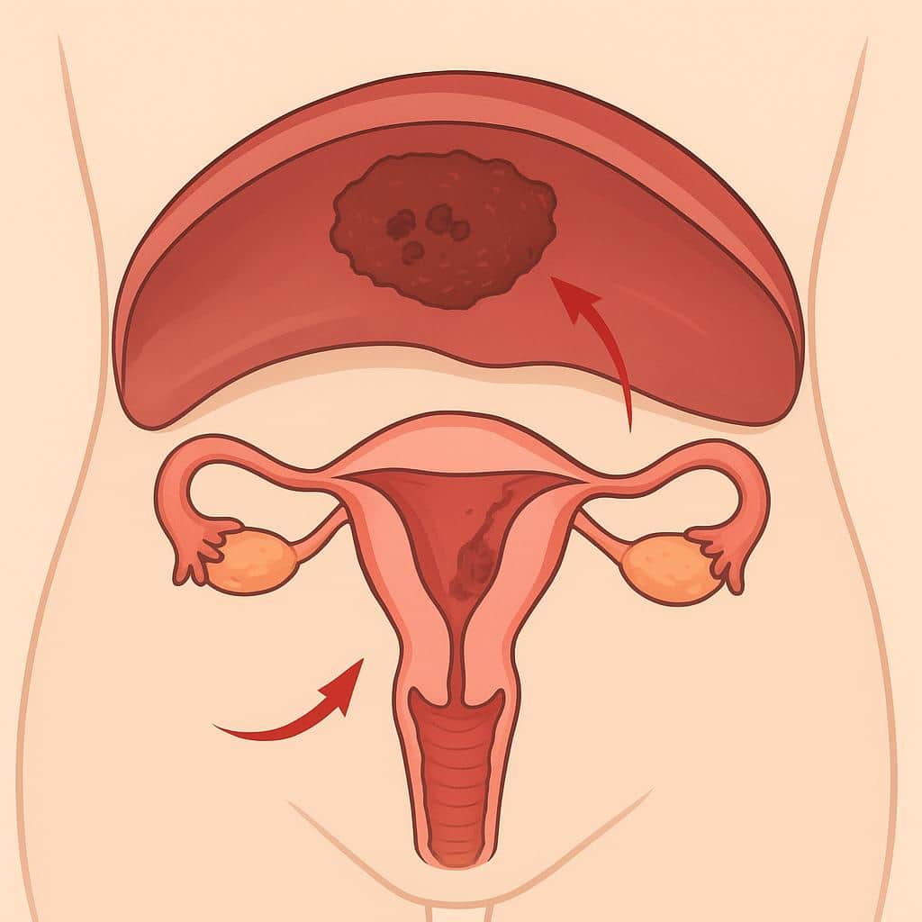
Diaphragmatic Endometriosis: Symptoms and Treatment Options
Learn what diaphragmatic endometriosis is, how it affects the diaphragm, key symptoms, causes, diagnosis options, treatments, and potential complications.
4 minute read
Read moreDiscover how Lotus can guide you toward lasting relief.
Explore why patients choose Lotus
Search expert-written answers, browse by topic, or find information based on where you are in your journey.
Explore All KnowledgeReach out and start your healing journey.
Schedule an AppointmentEvidence-based guides to how MRI detects and maps endometriosis, when it’s recommended, prep tips, and interpreting reports. Includes accuracy vs ultrasound and how findings inform treatment decisions.
MRI offers a detailed, radiation‑free way to detect and map endometriosis, especially deep infiltrating disease involving the bowel, bladder, ureters, or nerves. It is excellent for confirming ovarian endometriomas and assessing organ involvement, but it is less sensitive for superficial peritoneal lesions or subtle adhesions. Quality depends on protocol and expertise; centers may use antispasmodic medication and small amounts of vaginal or rectal gel to sharpen images. For adenomyosis‑specific imaging guidance, see Imaging & Diagnosis (MRI, Ultrasound).
MRI is typically recommended when expert Ultrasound is inconclusive, symptoms suggest complex or multi‑organ disease, or detailed mapping is needed before surgery. Results help tailor treatment and plan precise, organ‑sparing procedures alongside Imaging for Surgery, particularly for suspected Deep Infiltrating Endometriosis. Expect a 30–45 minute scan, loud sounds with ear protection, and usually no contrast; gadolinium is reserved for select questions. MRI can reduce the need for purely diagnostic Laparoscopy while clarifying whether medical or surgical approaches are most appropriate.
Yes. A ruptured ovarian cyst can cause sudden, severe pelvic pain, often sharp and one-sided, and may be intense enough to feel alarming, especially if there’s internal bleeding or irritation of the lining of the pelvis. Some people also notice nausea, shoulder-tip pain, dizziness, or pain that worsens with movement, while others have a milder ache that fades over hours to days.
Because pelvic pain has many look-alikes and coexisting causes (including endometriosis, adenomyosis, ovarian/paraovarian cysts, torsion, bladder pain, or pelvic floor spasm), what matters is the pattern of your symptoms, your exam, and correctly interpreted imaging such as ultrasound or MRI when appropriate. Our team focuses on sorting out whether a cyst rupture is the whole story or one piece of a bigger picture so you’re not stuck treating the wrong problem. If you’re having severe pain, recurrent episodes attributed to cysts, or pain that tracks with your cycle, reach out to schedule an evaluation so we can pinpoint the driver and map out next steps.
Imaging misses endometriosis more often than most patients are led to believe. Ultrasound and MRI can be very helpful for suggesting endometriosis and mapping certain patterns of disease (like ovarian endometriomas or some forms of deep disease), but a normal scan does not rule out endometriosis, especially superficial peritoneal disease, subtle lesions, or disease hidden in locations that are difficult to visualize.
How often imaging misses it depends on the type of endometriosis you have, where it is, the scan protocol, and, crucially, the experience of the person performing and interpreting the study. It’s also common for one modality to detect something the other doesn’t, because each has different blind spots (for example, bowel gas can limit MRI in some situations, while ultrasound may miss certain deep sites). That’s why our diagnostic process doesn’t hinge on imaging alone. We combine your symptom story, exam findings, and expert imaging interpretation to decide what’s most likely and what the next step should be.
If your symptoms persist despite negative imaging, that result is a data point, not a dead end. Reach out to schedule a consultation so our team can review your history and prior imaging, consider other conditions that can mimic or coexist with endometriosis, and determine whether advanced imaging, further evaluation, or surgical assessment makes sense for your goals.
Yes. It’s very possible to have endometriosis even when an ultrasound and MRI are read as normal, because imaging is not a simple yes-or-no test for all lesion types or locations. Scans are best at spotting certain patterns, like ovarian endometriomas or some deep disease, but superficial implants, small lesions, or disease hidden in harder-to-see areas can be missed. That’s why a normal scan should never automatically lead to dismissing symptoms like cyclical pelvic pain, painful sex, bowel/bladder symptoms, or infertility.
In our evaluation process, we use imaging as one part of the assessment, often more for mapping suspected disease and planning safe surgery than for ruling endometriosis in or out. We give considerable weight to your full symptom history, flare patterns, and a careful exam. We also look for conditions that can mimic or coexist with endometriosis (including adenomyosis, pelvic floor dysfunction, vascular causes of pelvic pain, and other drivers of inflammation). If your symptoms persist despite normal imaging, reach out to our team: our job is to bring these findings together and build a clear, actionable plan toward diagnosis and lasting relief.
Sciatic endometriosis is diagnosed by bringing together the symptom pattern, exam findings, and expert interpretation of imaging, then confirming the diagnosis when indicated. We start by listening closely to your full story, including whether buttock, back, or leg pain flares around your cycle, how far it radiates, and whether you’ve had numbness or tingling, weakness, gait changes, or foot drop. On exam, we look for findings that map to the sciatic nerve distribution. The exam can include maneuvers such as a straight-leg raise (Lasègue’s test) and assessment for deep tenderness near the sciatic notch.
Lab tests generally don’t diagnose sciatic endometriosis; inflammatory markers and sometimes CA-125 can be elevated but aren’t specific and don’t prove nerve involvement. MRI is often the most useful imaging tool for suspected endometriosis-related extraspinal sciatica because it may show a lesion along the nerve (commonly near the sciatic notch) or patterns of indirect compression or inflammation that can mimic piriformis syndrome. Even with good imaging, findings can be subtle, so symptoms outside the uterus or pelvis shouldn’t be dismissed. The diagnosis often depends on a careful, whole-body differential that also considers look-alike or coexisting causes of sciatica.
If your history and imaging raise concern for sciatic involvement, our team can guide a stepwise evaluation and discuss what confirmation and treatment would involve in your specific case, including when minimally invasive excision is appropriate and how we assess other contributors to persistent pain. If you’re experiencing progressive weakness, walking difficulty, or foot drop, we consider that a more serious presentation and prioritize timely assessment to reduce the risk of long-term nerve injury.
Hormonal birth control can absolutely make endometriosis harder to suspect on imaging, not because it reliably erases disease, but because it can quiet the inflammation and bleeding that make findings more obvious. Many endometriosis lesions (especially superficial disease) aren’t consistently visible on ultrasound or MRI to begin with, so symptom suppression can create a false sense of calm, with scans looking normal even when endometriosis is still present.
That said, imaging interpreted by an expert can still identify certain patterns (like ovarian endometriomas, deep infiltrating disease, adhesions, and related conditions such as adenomyosis) even if you’re on hormones. When we evaluate you, we don’t rely on imaging alone; we combine your symptom history and flare patterns with a careful exam and targeted imaging review to distinguish endometriosis from look-alike or coexisting causes of pelvic pain. If you’re worried that birth control is masking what’s going on, reach out to our team. We can help map out a diagnostic plan that doesn’t depend on a single test.
Diaphragmatic endometriosis can be difficult to confirm because symptoms may be subtle or absent, and imaging doesn’t always detect superficial implants. We start with your full symptom history and patterns, especially cyclical right upper abdominal, rib, chest, shoulder, or arm pain that flares around your period or with deep breaths or coughing. We then pair that with a targeted exam and a careful review of prior workups so we don’t miss look-alike or coexisting conditions.
Imaging such as MRI, and sometimes CT depending on the situation, can help raise suspicion, map anatomy, and guide surgical planning, but a normal scan does not rule it out. The most reliable way to diagnose diaphragmatic endometriosis is minimally invasive surgery (laparoscopy or robotic surgery) with deliberate inspection of the diaphragm and, when appropriate, removal of suspicious lesions for confirmation by pathology.
If symptoms suggest disease may extend into the chest (thoracic endometriosis), diagnosis may require coordination with a thoracic surgeon and, in select cases, a chest procedure such as VATS in addition to laparoscopy. Our team plans for this proactively when your history or imaging points in that direction, so you’re not left with an incomplete evaluation or surgery that isn’t equipped to address the full extent of disease.
“Junctional zone thickening” on MRI means the inner muscle layer of the uterus (the junctional zone, right next to the uterine lining) looks thicker and often less uniform than expected. This finding is commonly associated with adenomyosis, a condition where endometrial-like tissue grows into the uterine muscle, or myometrium, and can drive inflammation and pain.
It’s important to know that junctional zone thickening is not a definitive diagnosis by itself. It’s an imaging clue that needs to be interpreted alongside your symptoms (such as painful periods, heavy bleeding, pelvic pain, or fertility challenges) and the rest of the MRI details. Sometimes thickening can be more pronounced in one area, suggesting focal adenomyosis/adenomyoma. Adenomyosis can also overlap with endometriosis, which can change the overall plan.
If your report mentions junctional zone thickening, our team can help you understand the exact wording and what it likely means for you: whether it supports adenomyosis, whether the pattern looks focal or diffuse, and what next steps make sense based on your goals (symptom relief, fertility, or both). Reach out to schedule a consultation so we can review your imaging and history together.
Follow-up imaging doesn’t follow a single fixed schedule. It’s individualized based on what we’re monitoring. We use ultrasound and/or MRI most often when it will change decisions, such as tracking ovarian endometriomas, mapping suspected deep disease (bowel, bladder, or uterosacral involvement), or evaluating adenomyosis or a new pelvic mass. Symptoms and imaging don’t always match, so the goal is to get the right test at the right moment rather than scan on a timer.
In many cases, a practical framework is a baseline post-op check once initial healing is complete (often around 6–12 weeks), a planned reassessment around 6–12 months, and then annual follow-ups, especially if you had endometriomas, deep infiltrating disease, or persistent symptoms. Imaging may not be needed at every visit, but we’re more likely to recommend it when symptoms change, when prior imaging was incomplete, or when we need a clearer multi-compartment map to guide next steps. If you share your prior imaging reports, along with images if available, our team can tell you what’s worth repeating, what’s not, and how often surveillance makes sense for your specific pattern and goals.
Learn what diaphragmatic endometriosis is, how it affects the diaphragm, key symptoms, causes, diagnosis options, treatments, and potential complications.
Lotus Endometriosis Institute provides California-based surgical evaluation and advanced excision care for patients with suspected endometriosis, adenomyosis, complex pelvic pain, and related conditions.
Many patients contact us from outside California to learn whether traveling for in-person evaluation and possible surgery may be appropriate.
2121 Santa Monica Blvd, Santa Monica, CA 90404
8am - 5pm
Monday - Friday
154 Traffic Way, Arroyo Grande, CA 93420